A

David

Darling

hypodermis

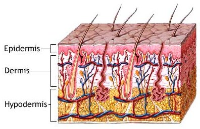
layers of the vertberate skin

Hypoglossal nerve, cervical plexus, and their branches Source: Gray's Anatomy (1918).


The hypodermis is a layer of tissue that lies immediately below the dermis of vertebrate skin. It is often referred to as subcutaneous tissue though this is a less precise and anatomically inaccurate term. The hypodermis consists primarily of loose connective tissue and lobules of fat. It contains larger blood vessels and nerves than those found in the dermis.


Specifically, the hypodermis contains:


  • Loosely arranged elastic fibers
  • Fibrous bands anchoring the skin to the deep fascia
  • Fat, except in the eyelid, scrotum, penis, nipple and areola
  • Blood vessels on route to the dermis
  • Lymphatic vessels on route from dermis
  • Hair follicle roots
  • The glandular part of some sudiferous glands
  • Nerves: free endings and Panicinian corpuscles
  • Bursae, in the space overlying joints in order to facilitate smooth passage of overlying skin
  • Fine, flat sheets of muscle, in certain locations, including the scalp, face, hand, nipple, and scrotuim, called the panniculus carnosus
  • In some animals, such as whales and hibernating mammals, the hypodermis forms an important insulating layer and/or food store.

    In some plants, the hypodermis is a layer of cells immediately below the epidermis of leaves. It is often mechanically strengthened, for example, in pine leaves, forming an extra protective layer or a water storage tissue.